A
PHYSICIST'S
VIEW OF MAGNETICS
BEING USED FOR
TREATING
MICROBIAL ASSOCIATED DISEASES
AND INFECTIONS
by Gary
Wade, Physicist
Editor’s Note: The interview was done in the interests of
clarification for those of us who are lay
persons and are interested in knowing why and how magnetic fields affect the body. For more information on pulsed magnetic therapy please see
Source Article
CB: You say that magnets affect the water molecules
in the blood plasma causing them to vibrate.
GW:
Yes, the ultra sound generated by interaction of the
moving blood plasma across the magnetic field can destroy a microbe.
CB: Do you mean then that microbes are the cause of
many of our illnesses?
GW: Yes, since microbes are inherently part of
life, an imbalance or overgrowth in any one of the microbial families can cause
havoc in human and animal bodies.
CB: Is it necessary to project different
levels of intensity for different forms of microbes?
GW: Yes, there are various forms of microbes that
respond to individual vibrational levels from low to
high.
CB: When we talk about microbes you mean, of
course, viruses, bacterias and fungi.
GW: Yes, plus protozoa and rickettsias—these
are the main cuprits I’d say.
CB: Are we correct to imagine that as the blood plasma moves across the magnetic field, oscillation is
created which ruptures the interlocking protein
clump rings, thereby rupturing the viruse’s outer
coat, which is known as the capsid?
GW: Yes, that’s essentially correct. However, it should be noted that every virus
has its own particular set of frequencies of vibration that destroys its capsid.
CB: Why do some practitioners of the healing
magnetic art use the positive side and other the negative side of the magnetic
field?
GW: Their claims seem to be based on empirical
results. However, with further research
by scientists, I suspect that a difference in the way enzymes work in the
presence of a north or south pole will be
observed. This is all speculative
because the research has not yet been done.
CB: Do you think its
important for people to become self sufficient to the greatest extent possible
with regard to their wellness/
GW: Absolutely.
Orthodox medicine is commonly 10-50 years behind in implementation of
research results and the pharmaceutical/FDA/AMA type organizations must
suppress technologies that threaten the profit centers of the status quo. After all, sickness care is big business.
For some years now new age health
practitioners have been using permanent magnets for the treatment of various
microbial associated afflictions. They
have been making some rather strong claims of successful treatment. When I have questioned such practitioners of
magnet therapy they invariably can not supply any technical or accepted
scientific reason for their apparent empirical treatment successes. In this article I will explain some aspects
of how and why the treatment works using
standard physics results in concert with the experimental research work of Dr.
Royal Raymond Rife.(1,2) The
short parcel answer to why permanent
magnet therapy works has two parts.
First, the arterial and veinal blood flow in
the presence of a magnetic field causes the generation of broad band ultrasound
by water molecules and ions in the blood plasma. Second, Dr. Rife found that each microbe type
has its own specific frequency of ultrasound which will destroy it.(3) Furthermore, the required intensity of the
ultrasound to destroy the microbe at the required specific frequency for that
microbe of interest, is ultra ultra low ( ~ 10-16 watts / meter squared ).(4) In other words the ultrasound generated by
the interaction of the magnetic field with the person's blood flow can destroy
a microbe, if the ultrasound frequency that the microbe is susceptible to is
one of the frequencies of ultrasound generated and is at a sufficient intensity
level.
Figure 1
illustrates the magnetic field of a flat wafer shaped dipole permanent
magnet. The magnetic field is invisible
and only manifests itself by its effects on electric current flow and
magnetically effected materials.
However, we are visually oriented creatures and like to visualize things
in order to think about them. Fortunately
the magnetic field
associate with common dipole permanent magnets allows for a easy
visual representation. You can
"see" the magnetic field by the use of diagrams which use the concept
of lines of flux. Such a diagram is
shown in Figure 1,
for a simple dipole wafer shaped permanent magnet. Figure 1 shows a cross-sectional view of the
flux lines for a plane cut through the middle of the dipole wafer magnet. The significance of the lines of flux diagram
is in the spacing distance between the adjacent lines. The larger the spacing, the
weaker the magnetic field strength.
The relative strength of the magnetic field at points a, b, and c in Figure 1 is the same as
the relative distances between the flux lines
shown. The direction of the magnetic
field at a point is parallel to the tangent of the flux line at that
point. Figure 2 shows a dipole
wafer permanent magnet placed on the "patient's" skin. The nature of the human blood circulatory
system is such that with a one inch in diameter wafer magnet there are dozens
to hundred of arterioles and veinules within a
quarter inch depth under the skin/wafer surface. In other words there are plenty of potential
ultra sound source zones over the entire body surface. To understand how relative motion between the
magnetic field and water molecules or ions in the blood flow can generate
ultrasound we need to see how charged particles react with mixed electric and
magnetic fields. Figure 3 shows the
motion of both a positive and a negative charged particle in a uniform electric
and magnetic fields which are at right angles to each other. Though the physic theory and mathematics
behind the goings on in Figure 3 are beyond most readers
educational experience, the results are simple to understand when
explained. I will explain Figure 3 and hopefully
verify my above statement for the laymen.
The uniformly spaced circles with X's in them ( lines
of flux or field lines ) represent a uniform magnetic field going directly into
the plane of the page. For example the
type of magnetic field you would have if the flat face of the north pole of the
dipole wafer permanent magnet were laying on the page
at that location. The uniformly spaced
parallel arrows in the plane of the page labeled E ,
which are at right angles to the magnetic field, are representative of an
electric field. Electric fields like
magnetic fields are invisible, but similar to magnetic fields can be visually
represented by electric field lines.
When the electric field lines are uniformly spaced and parallel as they
are in Figure 3, this
represents a uniform electric field of constant strength. Such an electric field can be generated
between two parallel metal plates, where the two plates have been given equal
magnitude net charges, one of a negative charge ( excess electrons ) and the
other a positive charge ( electron deficiency ). Now it is an experimental fact and a
mathematically derivable conclusion that both positively and negatively charged
particles will execute the type of motion depicted in Figure 3, when placed in
the type of crossed electric and magnetic fields depicted in Figure 3. Furthermore, we see that the charged
particles have a repetitive or cyclic motion.
In fact they have a well defined frequency of repetitiveness given by:
F = ( Q
) ( B ) / ( 2---) ( M ) ; Equation 1
where F is the number of half
circle arcs executed by the charged particle per second , Q is the magnitude of
the charge on the particle ( ion ), B is the absolute value of the magnetic field strength, and M is the
mass of the charged particle. The
charged particles depicted in Figure 3 are free to move in their cyclic motion
without collisions / interactions with other particles. This is not the case in general for ions in blood
plasma. Because of the extreme
compactness of the molecules in blood plasma, which consists mainly of water, the cyclic motion depicted in Figure 3 can only occur when the cyclic repeat
distance L of the
ion of Figure 3 is approximately half
the diameter of the blood plasma ion or smaller. To make ultrasound in blood plasma by this
cyclic motion we need weak interaction between the ion and other blood plasma
molecules. Detailed calculations for L where only weak interaction with other
molecules occur as applies to ions in blood plasma flowing in the presence of a
magnetic field gives:
L = ( C V sin w ) / B ; Equation
2
where V is the magnitude of the velocity of the
blood plasma flow relative to the magnetic field B, w is the angle between the direction of the
blood flow and the direction of the magnetic field and C is a
constant ( C = (2--- M) / 80 Q ). Now
looking back at Figure 2 we see that B,
V and w can take on a large combination of continuous sets of values
depending on just where in the blood flow system we are looking (
0< sin w <1 , 0 < V
< Vmax
). What equation 2
tells us is: Go to any point in
the blood plasma flow, and the values of
B, C, V, and w at that point determine the size (L) of the ion cyclic
displacement as it produces ultrasound of mainly a frequency given by equation
1. As long as the given L from equation 2
is smaller than approximately
half the ion's diameter , the ion will be an effective ultrasound
producer. Human blood plasma contains
eight major ion types, which can produce ultrasound when exposed to a magnetic field.
Now imagine that the positive and negative
particles in Figure 3
are elastically coupled together so that they can not escape from each
other. You now have what is known as a
charge dipole. As long as the charges
are not rigidly coupled together they will still have a tendency to execute the cyclic
motion, only now there will be more stringent constraints on the amplitude of
oscillation (" half circle" displacement ). Well just such a charge dipole is a water
molecule. A water molecule contains two
hydrogen atoms and one oxygen atom. The
oxygen atom has the two electrons associated with the hydrogen atoms in it
immediate vicinity 80+ % plus of the time.
This "hogging" of the
hydrogen electrons makes the oxygen atom effectively negative in charge and
leaves the hydrogen atoms deficit in electron charge and therefor
effectively positive in charge. The
water molecule however is a quantum mechanical system and can only exhibit
discrete ( quantized) oscillation modes. If a
isolated water molecule is placed in a crossed electric and magnetic
field as shown in Figure
3, the water molecule will
produce oscillatory motions of the
hydrogen and oxygen atoms only at quantized values and only at relatively very
high values for both the magnetic and electric fields. If the water molecule is in liquid water it can still only execute
quantized oscillation modes, however now the available number of oscillation
modes are very large, very closely spaced together in energy values, and
effectively start at zero frequency and zero energy. The reason for the change in available
oscillation modes ( states) is that water on an ultra
microscopic size scale behaves like a crystalline solid. Crystalline solids have phonons, which are quanitized vibration modes of the crystal
. These quantized vibrations
travel through the crystal structure at the speed of sound for that particular crystal. Phonons can be thought of as quantized pieces
of sound. The crossed electric and
magnetic field of Figure
3 can now couple their field energy into the production of phonons in
liquid water. The formula (Equation 1)
for the oscillation frequencies of free hydrogen and free oxygen atomic ions is
to a good first approximation the same as that for the maximum frequencies of
the phonons generated in the water by the crossed electric and magnetic
fields. The reader may now be wondering
where did the electric field used in Figure 3 to explain how ultrasound could
be generated in a situation as depicted in Figure 2 come
from. The answer is,
it is an experimental fact that when you move relative to and at an angle to the
direction of a stationary magnetic field, you experience an electric field
along with the magnetic field.
Furthermore, this induced electric field is at right angles to both your
velocity direction and the direction of the magnetic field. In other words the relative motion to a
stationary magnetic field generates an associated electric field at right
angles to the magnetic field just as depicted in Figure 3. Therefor ions and
water molecules being carried by the blood flow and moving relative to the stationary
magnetic field in Figure 2 experience a crossed electric
and magnetic field as depicted in Figure 3. Both the hydrogen and oxygen atoms, which have
an effective net charge in the water molecule execute quanitized bursts of cyclic motion. When these quanitized
bursts of cyclic motion take place in a highly compact and physically connected
medium such as water or blood plasma, it produces ultrasound.
To get some idea of the ultrasound
frequency range generated by this process, let M be the mass of a proton ( essentially all the mass of an hydrogen atom ), Q be 80 %
the charge of an electron , but of a positive sign, and B be .2 webers/meter squared at the magnet pole face, a common
value for strong ceramic magnets.
Putting these values into Equation 1, we obtain for the maximum
ultrasound frequency, 2.54 X 10⁶ cycles per second. Now note as you go away from the magnet pole
face ( going deeper into the body ) the magnetic field strength drops off
quickly, and therefor so does the frequency of
ultrasound generated since we see from Equation 1 that the frequency is
directly proportional to the strength of the magnetic field. In other words a continuum of ultrasound frequencies
starting at the maximum frequency and going to zero frequency is generated in
the body tissue under the magnet pole face.
Of coarse we could have chosen a permanent magnet or an electro-magnet
which is the state of the art of technology and achieved several times higher
maximum frequency for the continuum of ultrasound frequencies generated.
To see how the ultrasound generated by the
interaction of a magnetic field with blood flow can destroy a virus we will
examine the outer protein coat ( capsid
) structure of a virus. Most viruses of
interest which cause diseases in plants and animals have an icosahedral
capsid structure as illustrated in Figure 4A and B. A specific example of this icosahedral
capsid structure is illustrated in Figure 5. Each dark circle represents a spherical
protein molecule clump. When the virus capsid of Figure 5 is folded
together as indicated in Figure
4A and B, a simple virus capsid model has been
formed. Examination of this capsid model shows a large number of intersecting and
overlapping closed rings of protein molecule clumps. These closed rings of periodically spaced
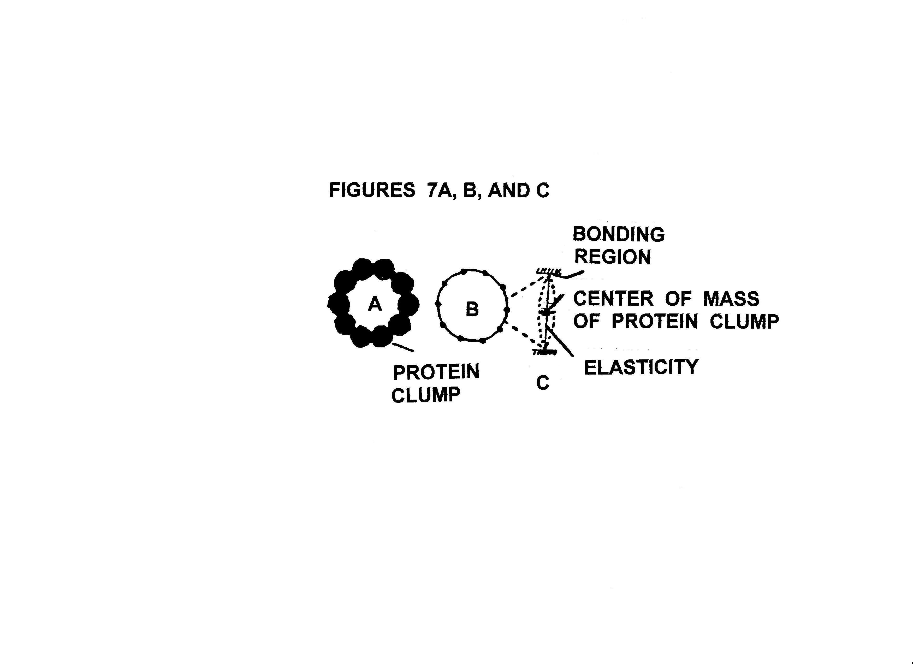
protein clumps are illustrated in Figure 6A,B,C, and D. In
classical physics when studying standing wave phenomenon the periodically
spaced protein clumps as illustrated in Figure 6A,B,C, and D
are known as the mass beads on a string problem with circular boundary
conditions. Figures 7A,B, and C illustrate this classical physics problem for
the ten member protein clump ring. Figures 8A and B illustrate one of the standing wave motion modes which the
closed periodically spaced protein clump ring of Figures 6A can sustain. Figure 8A shows a ten member
protein clump ring linearized for ease of graphing
wave motion displacement of the center of the protein clumps from their
equilibrium position. Figure 8B shows the most
stressful oscillation mode for the ten member protein clump ring. In this oscillation mode adjacent protein
molecule clumps are always going in opposite directions and therefor
putting maximum stress on where they are bonded together. If this oscillation mode is raised to a high
enough displacement amplitude the ring will rupture. If enough rings are ruptured, the virus capsid disintegrates and the virus is destroyed..
It is important to construct a virus capsid model to get a full and clear appreciation of how
susceptible a virus is to ultrasound of its resonance frequency(ies). Here are the
instructions for assembly of the virus coat in Figure 5. First go to a copy machine and enlarge the
figure 122% two or three times. This
will give almost a page full of virus coat.
Second, make another copy of the last enlargement onto thin cardboard
copy paper. You may have to look around
to find the right copy machine for this.
Third, cut out all dashed lines on the cardboard copy. You should now have something that looks like
Figure 4A except
for extra tabs for gluing the model together. I have found that Elmer's Glue All works
well. Fourth, taking a straight edge
ruler and lining its edge up congruent with all of the equilateral triangle
facet edges as those shown in Figure 4A, fold the cardboard over the ruler edge
until a 90 degree fold angle is achieved, while folding away from the faces
shown in Figure 5. Be sure to fold all of the glue tabs this way
also. Fifth, begin gluing adjacent tabs flush
together. It may prove helpful to use
scotch tape to tape the adjacent faces together after gluing while waiting for
the glue to set. Also strong alligator
clips are useful in holding the glued tabs together while the glue sets up. Have fun and may the glue be with you.
The common virus capsid
coat was chosen to show how ultrasound generated standing waves can destroy a
microbe which has closed on themselves periodically spaced protein clump
structures. Bacteria, protozoa, rickettias, and fungi all have these closed on themselves
periodically spaced protein clump structures in their outer structure which
makes them susceptible to destruction by ultrasound. In our present circumstances where antibiotic
resistant bacteria are about to become rampant, and anti-viral drugs are largely still
just a bio-tech dream, it is time to know of ways to treat yourself, if the
medical establishment has no viable answers
or you must wait until you die while the FDA gets around to approving a
viable treatment, which has been known and well documented for many years by
honest competent researchers.
Advancements in electronics have made a
much more efficient and vastly more powerful replacement for the permanent
magnet method of ultrasound
generation to kill microbes.
Namely, the piezo-electric transducer, driven
by an appropriate signal function generator as can be purchased in any
electronic test equipment store. By
merely applying a triangle voltage wave form from the function generator to the
piezo-electric transducer, pressure square waves are
produced by the transducer. These
pressure square waves have effectively hidden in them an infinite set of sine
wave pressure waves of frequencies equal to odd integer multiples of the
frequency of the triangle voltage wave.
By very slowly varying the frequency of the triangle voltage wave form
from its lowest to its highest value on the function generator, a set of
pressure sine waves is generated by the transducer , which covers a frequency
range of two hundred thousand to twenty million plus cycles per second for most
commonly available function generators.
This ultrasound frequency range is sufficient to kill the vast majority
of viruses, bacteria, protozoa, fungi, and rickettias
known. With most function generators the
application of a very slowly varying o to +10 volts or 0 to -10 volts to the voltage controlled
frequency ( VCF) input will vary the triangle voltage wave frequency over its
entire range.
There are still many reported empirical
effects / results of the new age healers using dipole permenate
magnets, which I have not been able to verify or explain yet. However, I feel confident that once
scientists begin to take these effects / reported empirical results seriously
and then do serious experiments / calculations on the effects of strong
magnetic fields from dipole permenate magnets on
enzyme chemistry and cell physiology, that many of these effects / results will
be put on strong theoretical ground. One
of the most interesting reported empirical results is that the magnetic north
pole must be face down on the skin over the infected area for the treatment of
infections and cancer to work.
Furthermore, if the other magnetic pole ( south
pole ) is face down on the skin it supposedly often worsens the condition. However, when I tested a magnet used by a new
age healer at a recent health show I found that he was treating infections with
the south magnetic pole down on the skin.
So, I find this aspect a point of confusion needing resolution.
One point I would like this article to
make is that just because some authority figure or organization or government bureaucracy says that some treatment or product or whatever
is of no value because there is no scientific evidence to support the reported
empirical results, it does not matter, their opinion when based on ignorance,
self / vested interest, and stupidity is no more valid than yours or mine when
our opinions are based on a similar foundation. It should also be clearly noted that
authority figures, government bureaucrats, organization PR personnel all
commonly lie to the general public. And
in my opinion this is something we should begin to punish them serverly for doing.
Perhaps, knowingly lying to the public about a substantive issue should
be made a mistimeaner that carries a mandatory ten
day jail term plus a stiff fine.
References:
1) A Physicist's View Of
Dr. Rife's Non Drug Treatment And Cure Of Microbial Associated Diseases by Gary Wade,
Health Freedom News, August 94
2) The Royal R. Rife Report compiled by Alison Dividson, published by Borderland Sciences,
3) What Has Become Of The Rife
Microscope? by Christopher Bird, New Age Journal,
March 1976.
4) Dr. Rife And The Death Of The Cancer Industry
( see Appendix B ) by Gary Wade, self published, for info right Gary Wade, P.O.
Box 3813, Alhambra, CA. 91803
IF YOU FOUND THIS ARTICLE OF REAL VALUE, PLEASE MAKE A HARD COPY WHILE STILL AVAILABLE.
BACK TO PULSED MAGNETIC
ARTICLE